
Making cancer vaccines more personal
Researchers used modeling to better understand which tumor proteins can provoke a powerful immune system attack against a type of skin cancer.
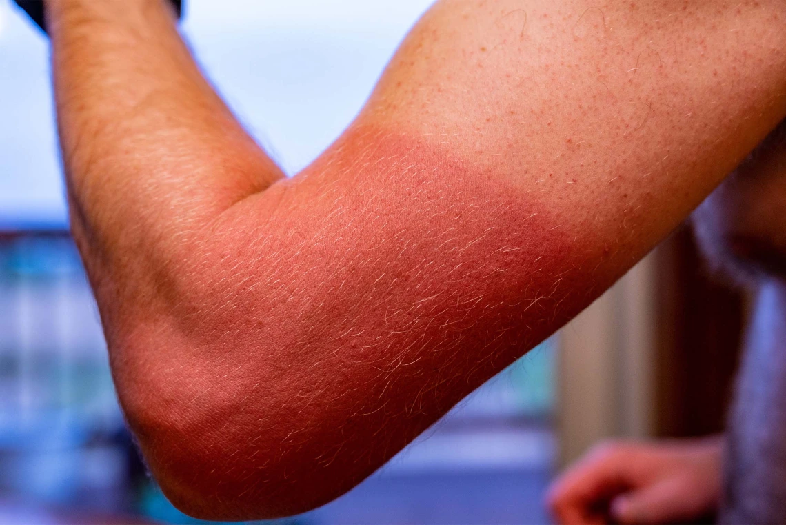
Prolonged sun exposure can lead to the development of cutaneous squamous cell carcinoma, a skin cancer that College of Medicine – Phoenix researchers are working to prevent.
Photo by Lost_in_the_Midwest via Adobe Stock
In a new study, University of Arizona researchers created a model for cutaneous squamous cell carcinoma, a type of skin cancer, and identified two mutated tumor proteins, or neoantigens, that contain features of good candidates for a vaccine. At the same time, they used artificial intelligence to create 3D models to help them understand and predict which neoantigens could provoke T cells, a type of white blood cell critical to the immune system, to attack the cancer.
Tumor neoantigens are unique mutated proteins in cancer cells. They act like an alarm system, alerting the immune system that cancer cells are a threat. By identifying and characterizing neoantigens, researchers can develop personalized tumor vaccines to help the immune system recognize and attack cancer cells.
The results suggest that both the structural and physical features of neoantigens could play important roles in predicting which ones could be used in cancer vaccines against tumors. The findings were published in the Journal for ImmunoTherapy of Cancer.
Dr. Karen Taraszka Hastings is a professor and chair of the Department of Dermatology at the U of A College of Medicine-Phoenix.
Photo by Noelle Rosario Haro-Gomez, U of A Health Sciences Office of Communications
“One of the challenges in creating tumor-based cancer vaccines is finding the right mix of neoantigens to elicit a T cell response that can destroy a tumor,” said senior author Dr. Karen Taraszka Hastings, professor and chair of the Department of Dermatology at the University of Arizona College of Medicine – Phoenix and a member of the U of A Cancer Center. “We want to find ways to make it easier to choose the right neoantigens to include in cancer vaccines, especially in cancers like cutaneous squamous cell carcinoma and melanoma that contain a high number of mutations.”
Tumor vaccines can include dozens of mutated tumor protein fragments, or peptides. Some experimental vaccines aimed against mutations in a person’s tumor already exist, including for melanoma and pancreatic and non-small cell lung cancers. But for some cancers, such as cutaneous squamous cell carcinoma, there are thousands of tumor peptide mutations and no good way to figure out which mutations will be most useful in a vaccine.
The researchers found both human and mouse cancers have a high number of mutations, including the same key mutations that are instrumental in causing tumors. Using a mouse model, they also identified two neoantigens that prompted T cells to halt tumor growth.
Both of the neoantigens caused T cells to produce a strong anti-tumor response, while the normal version of the peptides did not. When they looked closer, they saw that each of the neoantigens worked differently though both were equally visible to the immune system.
Before T cells can act against tumor neoantigens, the immune system has to recognize them. The neoantigens must first attach to the major histocompatibility complex, or MHC, a set of proteins that act as a display case.
The team discovered that the MHC displayed the mutated Picalm peptide, whereas it didn’t display the normal peptide. This is likely responsible for the ability of mutated Picalm peptide to stimulate an anti-tumor T cell response. In contrast, the mutated Kars peptide and the normal peptide were bound to the MHC in similar ways.
“So, there is a different reason that mutated Kars elicits a T cell response that destroyed the tumor,” Hastings said.
To find out why, the scientists turned to 3D, artificial intelligence-based modeling to look for differences in the structures of the peptides and how they connected to the MHC.
“We found that the mutated Kars peptide has a different chemical structure on the surface of the 3D structure that is exposed to the T cell receptor,” Hastings said. “It is likely that this difference in the peptide structure between mutated and normal Kars is recognized by the T cell receptor, which results in a response that restricts tumor growth in mice.”
They examined all known cancer neoantigens that have been tested individually for the ability to control tumor growth and found that increased exposure of the mutated peptide to the T cell receptor was key.
“We are proposing 3D structural modeling as a way to further narrow down which neoantigens to select for inclusion in cancer vaccines, which should improve their effectiveness,” she said.
Hastings believes 3D modeling may be especially important for predicting useful neoantigens for developing individualized vaccines against skin cancers and melanoma, and it could have applications for other types of cancer as well. The team plans to test their ideas on human tumor samples next.
“The use of artificial intelligence in developing a new approach to building personalized cancer vaccines speaks to the transformative nature of the technology for cancer therapeutics,” said David Ebert, chief AI and data science officer at the U of A. “Dr. Hastings’ research is a perfect example of the potential impact of the University of Arizona on the future of medicine and patient care.”
Co-authors from the U of A College of Medicine – Phoenix include MD/PhD student Anngela Adams, Dr. Janko Nikolich, Regents Professor and Associate Dean of Research; research professional Anne Macy; MD/PhD student Elizabeth Borden; research technician Lauren Herrmann; and researcher/scientist Dr. Sandip Ashok Sonar. Additional U of A co-authors include Denise Roe, professor of biostatistics at the Mel and Enid Zuckerman College of Public Health. Also contributing to the study were Ken Buetow and Melissa Wilson of Arizona State University; Brian Baker of the University of Notre Dame; and Dr. Aaron Mangold of Mayo Clinic Arizona.
This work was supported in part by the National Cancer Institute, a division of the National Institutes of Health, under award numbers T32CA009213, F30CA257622, F30CA281056, P30CA023074 and P01CA229112, by the National Institute of General Medical Sciences, also a division of the NIH, under award number R35GM118166, and by an American Cancer Society – Simone Charitable Foundation - Discovery Boost Grant under award number AZ-DBG-23-1151311-01-IBCD.